20. Answer B is the correct answer. Torsades de point translated means "turning on a point." It literal looks like it is rotating around the point. Ventricular fibrillation is a fine wavy baseline and ventricular tachycardia is a wide complex tachycardia. Supraventricular tachycardia is a narrow complex tachycardia because its pacemaker is coming from outside of the ventricles.
Tuesday, December 24, 2013
Question 20
20. What is the diagnosis of the rhythm listed below:
 A. Ventricular Fibrillation
B. Torsades de Point
C. Supraventricular Tachycardia
D. Ventricular Tachycardia
B. Torsades de Point
C. Supraventricular Tachycardia
D. Ventricular Tachycardia
Answer 19
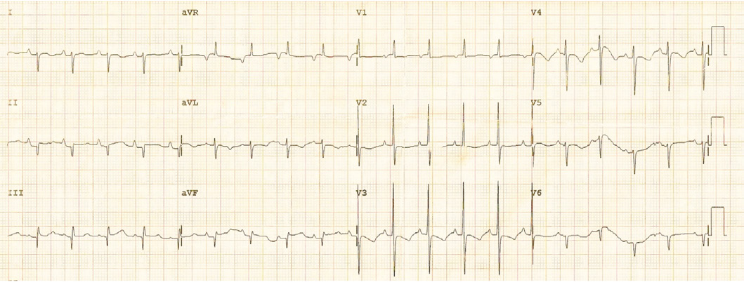
19. Answer B is the correct answer. With left ventricular hypertrophy, the QRS complexes become bigger in height and depth in leads V1-V6. The S wave will be even deeper in V1. The largest R wave will be in lead V5 because it lays over the left ventricle. The T wave will often be inverted or cause asymmetry. With right ventricular hypertrophy, there is large R wave in lead V1, there is amore positive deflection toward the V1 electrode and the QRS complex more upright than nomad. The R wave gets progressively smaller in leads V2-V6. With right atrial hypertrophy, the P wave is affected.
Question 19
19. Please identify the abnormality seen on the EKG below:

A. Right Ventricular Hypertrophy
B. Left Ventricular Hypertrophy
C. Right Atrial Hypertrophy
D. Normal EKG

A. Right Ventricular Hypertrophy
B. Left Ventricular Hypertrophy
C. Right Atrial Hypertrophy
D. Normal EKG
Answer 18
18. Answer D is the correct answer. With extreme right axis deviation, there is a negative deflection of the QRS complex is leads I and avF. With a right axis deviation, there is a negative deflection in lead I and a positive deflection in lead avF. With a normal axis, there is a positive deflection in leads I and avF. With a left axis deviation, there is a positive deflection in lead I and a negative deflection in lead avF.
Question 18
18. Please identify the pathology listed below on the EKG:

A. Normal Axis
B. Left Axis Deviation
C. Right Axis Deviation
D. Extreme Right Axis Deviation
A. Normal Axis
B. Left Axis Deviation
C. Right Axis Deviation
D. Extreme Right Axis Deviation
Answer 17
17. Answer B is the correct answer. With hypokalemia, the T wave becomes flattened, and there may be a U wave associated with it. With hyperkalemia, the P wave is flattened, the QRS complex is widened, and the T wave is peaked. With hypercalcemia, the QT interval is shortened. With hypocalcemia, the QT interval is lengthened.
Subscribe to:
Posts (Atom)